The dwarf planet Ceres -- long believed to be a barren space rock -- is an ocean world with reservoirs of sea water beneath its surface, the results of a major exploration mission showed Monday.
Ceres is the largest object in the asteroid belt between Mars and Jupiter and has its own gravity, enabling the NASA Dawn spacecraft to capture high-resolution images of its surface.
Now a team of scientists from the United States and Europe have analyzed images relayed from the orbiter, captured around 35 kilometers (22 miles) from the asteroid.
They focused on the 20-million-year-old Occator crater and determined that there is an "extensive reservoir" of brine beneath its surface.
Several studies published Monday in the journals Nature Astronomy, Nature Geoscience and Nature Communications also shed further light on the dwarf planet, which was discovered by Italian polymath Giuseppe Piazzi in 1801.
Using infrared imaging, one team discovered the presence of the compound hydrohalite -- a material common in sea ice but which until now had never been observed off of Earth.
Maria Cristina De Sanctis, from Rome's Istituto Nazionale di Astrofisica said hydrohalite was a clear sign Ceres' used to have sea water.
"We can now say that Ceres is a sort of ocean world, as are some of Saturn's and Jupiter's moons," she told AFP.
The team said the salt deposits looked like they had built up within the last two million years -- the blink of an eye in space time.
This suggests that the brine may still be ascending from the planet's interior, something De Sanctis said could have profound implications in future studies.
"The material found on Ceres is extremely important in terms of astrobiology," she said.
"We know that these minerals are all essential for the emergence of life."
Writing in an accompanying comment article, Julie Castillo-Rogez, from the California Institute of Technology's Jet Propulsion Laboratory, said the discovery of hydrohalite was a "smoking gun" for ongoing water activity.
"That material is unstable on Ceres' surface, and hence must have been emplaced very recently," she said.
In a separate paper, US-based researchers analyzed images of the Occator crater and found that its mounds and hills may have formed when water ejected by the impact of a meteor froze on the surface.
The authors said their findings showed that such water freezing processes "extend beyond Earth and Mars, and have been active on Ceres in the geologically recent past".
A new study reveals that male cicadas, when infected with a mind-controlling fungus that devours large parts of their bodies, will trick other male cicadas into being infested by the same horrifying parasite — thanks in part to a psychoactive chemical found in hallucinogenic mushrooms.
One of the chemicals in this fungus is psilocybin, the psychoactive that causes humans to "trip" when they consume psychedelic mushrooms. Intriguingly, scientists believe that in infected cicadas, this drug may actually help the cicada endure the trauma of having the lower half of its bottom replaced by said fungus while the upper half decays. This is important, as the fungus starts "pulling the behavioral levers" of the cicada through various chemical interactions with the insect's biology.
The researchers, including lead study author Brian Lovett from the Division of Plant and Soil Sciences at West Virginia University, describe how the fungus Massospora is able to engage in active host transmission, a process in which a pathogen uses "a form of biological puppetry" to manipulate one living host in order to transfer its material to another living host. This is why they are colloquially referred to as "zombie cicadas," both in the classical definition of that term (a dead or hypnotized being whose mind is controlled by another) and the contemporary cinematic one (a body that is literally decaying but continues to move around).
"Manipulation of a host to focus on pathogen transmission is fascinating, because it raises questions about the nature of autonomy and shines a light on the physical and behavioral manifestations of parasitism," the study, which was published in the journal PLOS Pathogens, says.
Salon reached out to Lovett to learn more about what this study suggests about the nature of free will in both insects and humans.
"We wouldn't claim to know what these cicadas are feeling or thinking during infection, but it's certainly tempting to empathize: how horrified would you be to discover that a sizable part of your body has been replaced by a fungus?" Lovett said by email. "What we can do is look at how some of the interesting compounds these fungi produce may affect the behavior of their cicada victims."
When describing periodical cicadas, Lovett observed that "an amphetamine (cathinone) is produced. This drug certainly changes our mood and behavior, and it has been shown to alter aggression and feeding in other insects. We speculate these fungal drugs may help cicadas endure the trauma of having their abdomen replaced by fungus, as they continue to spread its spores."
I asked Lovett if his research suggests that a similarly mind-controlling organism could rob human beings of their free will. Lovett laughed and replied, "I do not see a fungus evolving to replace our butts anytime soon. Modern medicine and pants prevent that from becoming a winning fungal lifestyle. However, there are already examples of parasites that manipulate our mood and behavior. In our PLOS Pathogens article, we highlight rabies as a pathogen that changes who we are to the benefit of the virus."
Indeed, humans who suffer rabies experience hydrophobia and difficulty swallowing, which lets the virus build up in the victims' mouth. They also become more aggressive, and in animal cases, will bite others, thus spreading the virus. In other words, rabies is a good example of a virus altering mammals' behavior in the virus' favor.
Massospora also has the dubious distinction of being the only known example of active host transmission in which "the pathogen behaves at least in part as a sexually transmitted disease, although natural history studies are lacking." Massospora often infects cicadas from the Magicicada genus, also known as periodical cicadas, which includes seven species that are known to spend either 13 or 17 years underground as nymphs before emerging for a few weeks to mate and die.
To find a partner, cicadas from this genus will practice "highly stereotyped" behavior in which "males call and females respond with wing flicks, but healthy males never signal with wing flicks. When females remain unmated much beyond the onset of sexual receptivity, their responses become exaggerated with louder, more consistent wing flicks and sometimes even whole-body motions that appear to draw the attention of chorusing males."
This is where Massospora enters the picture. The spores will force the zombified cicadas to move their wings in a way that other males of the species will associate with females seeking to mate. Once the unsuspecting male approaches the supposed female, the spores will be able to infect the new host. Instead of being able to mate with other cicadas and lay eggs before dying, the zombie cicadas are manipulated by the fungus to fly around so they can spread more spores.
In a visceral passage on why the term "zombie cicada" is visually appropriate, Nature's Scientific Reports wrote in 2018 that after copulation it is not uncommon "to see healthy cicadas attached to fragments of abdomen or terminalia that have torn free from infected partners during attempted copulation."
Anyone who’s tending a garden right now knows what extreme heat can do to plants. Heat is also a concern for an important form of underwater gardening: growing corals and “outplanting,” or transplanting them to restore damaged reefs.
The goal of outplanting is to aid coral reefs’ natural recovery process by growing new corals and moving them to the damaged areas. It’s the same idea as replanting forests that have been heavily logged, or depleted farm fields that once were prairie grasslands.
I have studied how global stressors such as ocean warming and acidification affect marine invertebrates for more than a decade. In a recently published study, I worked with Gregory Asner to analyze the impacts of temperature on coral reef restoration projects. Our results showed that climate change has raised sea surface temperatures close to a point that will make it very hard for outplanted corals to survive.
Rising ocean temperatures cause bleaching events – episodes in which corals expel the algae that live inside them and provide the corals with most of their food, as well as their vibrant colors. When corals lose their algae, they become less resistant to stressors such as disease and eventually may die.
Hundreds of organizations worldwide are working to restore damaged coral reefs by growing thousands of small coral fragments in nurseries, which may be onshore in laboratories or in the ocean near degraded reefs. Then scuba divers physically plant them at restoration sites.
Outplanting is the process of transplanting nursery-grown corals onto reefs.
Outplanting coral is expensive: According to one recent study, the median cost is about US$160,000 per acre, or $400,000 per hectare. It also is time-consuming, with scuba divers placing each outplanted coral by hand. So it’s important to maximize coral survival by choosing the best locations.
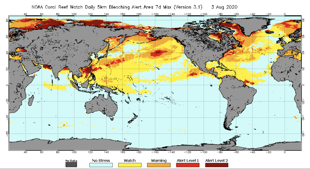
We used data from the National Oceanic and Atmosphere Administration’s Coral Reef Watch program, which collects daily satellite-derived measurements of sea surface temperature. We paired this information with survival rates from hundreds of coral outplanting projects worldwide.
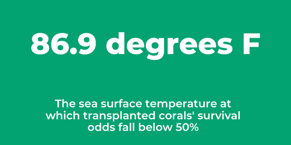
We found that coral survival was likely to drop below 50% if the maximum temperature experienced at the restoration site exceeded 86.9 degrees Fahrenheit (30.5 degrees Celsius). This temperature threshold mirrors the tolerance of natural coral reefs.
Globally, coral reefs experience an annual maximum temperature today of 84.9˚F (29.4˚C). This means they already are living close to their upper thermal limit.
When reefs experience temperatures only a few degrees above long-term averages for a few weeks, the stress can cause coral bleaching and mortality. Increases of just a few degrees above normal caused three mass bleaching events since 2016 that have devastated Australia’s Great Barrier Reef.
Sea surface temperatures on Aug. 3, 2020, measured from satellites. Warning = possible bleaching; Alert Level 1 = significant bleaching likely; Alert Level 2 = severe bleaching and significant mortality likely.
When coral restoration experts choose where to outplant, they typically consider what’s on the seafloor, algae that could smother coral, predators that eat coral and the presence of fish. Our study shows that using temperature data and other information collected remotely from airplanes and satellites could help to optimize this process. Remote sensing, which scientists have used to study coral reefs for almost 40 years, can provide information on much larger scales than water surveys.
Coral reefs face an uncertain future and may not recover naturally from human-caused climate change. Conserving them will require reducing greenhouse gas emissions, protecting key habitats and actively restoring reefs. I hope that our research on temperature will help increase coral outplant survival and restoration success.
These effects may be caused by direct viral infection of brain tissue. But growing evidence suggests additional indirect actions triggered via the virus’s infection of epithelial cells and the cardiovascular system, or through the immune system and inflammation, contribute to lasting neurological changes after COVID-19.
Many of the symptoms we attribute to an infection are really due to the protective responses of the immune system. A runny nose during a cold is not a direct effect of the virus, but a result of the immune system’s response to the cold virus. This is also true when it comes to feeling sick. The general malaise, tiredness, fever and social withdrawal are caused by activation of specialized immune cells in the brain, called neuroimmune cells, and signals in the brain.
These changes in brain and behavior, although annoying for our everyday lives, are highly adaptive and immensely beneficial. By resting, you allow the energy-demanding immune response to do its thing. A fever makes the body less hospitable to viruses and increases the efficiency of the immune system. Social withdrawal may help decrease spread of the virus.
Unfortunately, this also provides a way in which illnesses like COVID-19 can cause both acute neurological symptoms and long-lasting issues in the brain.
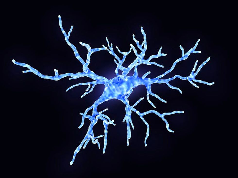
Microglia are specialized immune cells in the brain. In healthy states, they use their arms to test the environment. During an immune response, microglia change shape to engulf pathogens. But they can also damage neurons and their connections that store memory.
During illness and inflammation, the specialized immune cells in the brain become activated, spewing vast quantities of inflammatory signals, and modifying how they communicate with neurons. For one type of cell, microglia, this means changing shape, withdrawing the spindly arms and becoming blobby, mobile cells that envelop potential pathogens or cell debris in their path. But, in doing so, they also destroy and eat the neuronal connections that are so important for memory storage.
Another type of neuroimmune cell called an astrocyte, typically wraps around the connection between neurons during illness-evoked activation and dumps inflammatory signals on these junctions, effectively preventing the changes in connections between neurons that store memories.
Because COVID-19 involves a massive release of inflammatory signals, the impact of this disease on memory is particularly interesting to me. That is because there are both short-term effects on cognition (delirium), and the potential for long-lasting changes in memory, attention and cognition. There is also an increased risk for cognitive decline and dementia, including Alzheimer’s disease, during aging.
How does inflammation exert long-lasting effects on memory?
If activation of neuroimmune cells is limited to the duration of the illness, then how can inflammation cause long-lasting memory deficits or increase the risk of cognitive decline?
Both the brain and the immune system have specifically evolved to change as a consequence of experience, in order to neutralize danger and maximize survival. In the brain, changes in connections between neurons allows us to store memories and rapidly change behavior to escape threat, or seek food or social opportunities. The immune system has evolved to fine-tune the inflammatory response and antibody production against previously encountered pathogens.
Another major illness with a similar cognitive complications is sepsis – multi-organ dysfunction triggered by inflammation. In animal models of these diseases, we also see impairments of memory, and changes in neuroimmune and neuronal function that persist weeks and months after illness.
Does COVID-19 increase risk for cognitive decline?
It will be many years before we know whether the COVID-19 infection causes an increased risk for cognitive decline or Alzheimer’s disease. But this risk may be decreased or mitigated through prevention and treatment of COVID-19.
Prevention and treatment both rely on the ability to decrease the severity and duration of illness and inflammation. Intriguingly, very new research suggests that common vaccines, including the flu shot and pneumonia vaccines, may reduce risk for Alzheimer’s.
Additionally, several emerging treatments for COVID-19 are drugs that suppress excessive immune activationand inflammatory state. Potentially, these treatments will also reduce the impact of inflammation on the brain, and decrease the impact on long-term brain health.
COVID-19 will continue to impact health and well-being long after the pandemic is over. As such, it will be critical to continue to assess the effects of COVID-19 illness in vulnerability to later cognitive decline and dementias.
In doing so, researchers will likely gain critical new insight into the role of inflammation across the life-span in age-related cognitive decline. This will aid in the development of more effective strategies for prevention and treatment of these debilitating illnesses.
This year's Atlantic hurricane season could be one of the busiest on record, with as many as 25 named storms, forecasters said Thursday.
Due to atmospheric and oceanic conditions, "this year, we expect more, stronger, and longer-lived storms than average," said Gerry Bell, lead seasonal hurricane forecaster at the National Oceanographic and Atmospheric Administration's Climate Prediction Center.
Forecasters predict this season will see 19 to 25 named storms, of which seven to 11 will reach hurricane strength.
Of these, three to six will be major hurricanes, with winds of 111 miles per hour (180 kph) or greater.
The NOAA was updating a forecast from May that called for 13-19 named storms, with up to 10 of them growing into hurricanes.
The NOAA said this is one of the most active forecasts it has made in its 22-year history of predicting hurricanes.
The forecast includes the nine storms -- two of which became hurricanes -- that have formed so far in what the NOAA said could be an "extremely active" hurricane season.
People in the southern US and the Caribbean have already gotten a taste of violent weather in recent weeks.
Normally, at this time of year there have only been two named storms and the ninth does not come until early October. An average season has 12 named storms.
But Isaias, the ninth storm so far, just hit the Caribbean and the US east coast, leaving five dead as if drifted between hurricane and tropical storm status.
In late July, just days before Isaias, Category 1 Hurricane Hanna formed in the Gulf of Mexico and hit Texas, but without causing major damage.
So focus shifted to the relative sizes of brain regions. Phrenologists suggested the part of the cerebrum above the eyes, called the frontal lobe, is most important for intelligence and is proportionally larger in men, while the parietal lobe, just behind the frontal lobe, is proportionally larger in women. Later, neuroanatomists argued instead the parietal lobe is more important for intelligence and men’s are actually larger.
In the 20th and 21st centuries, researchers looked for distinctively female or male characteristics in smaller brain subdivisions. As a behavioral neurobiologist and author, I think this search is misguided because human brains are so varied.
Anatomical brain differences
The largest and most consistent brain sex difference has been found in the hypothalamus, a small structure that regulates reproductive physiology and behavior. At least one hypothalamic subdivision is larger in male rodents and humans.
But the goal for many researchers was to identify brain causes of supposed sex differences in thinking – not just reproductive physiology – and so attention turned to the large human cerebrum, which is responsible for intelligence.
Within the cerebrum, no region has received more attention in both race and sex difference research than the corpus callosum, a thick band of nerve fibers that carries signals between the two cerebral hemispheres.
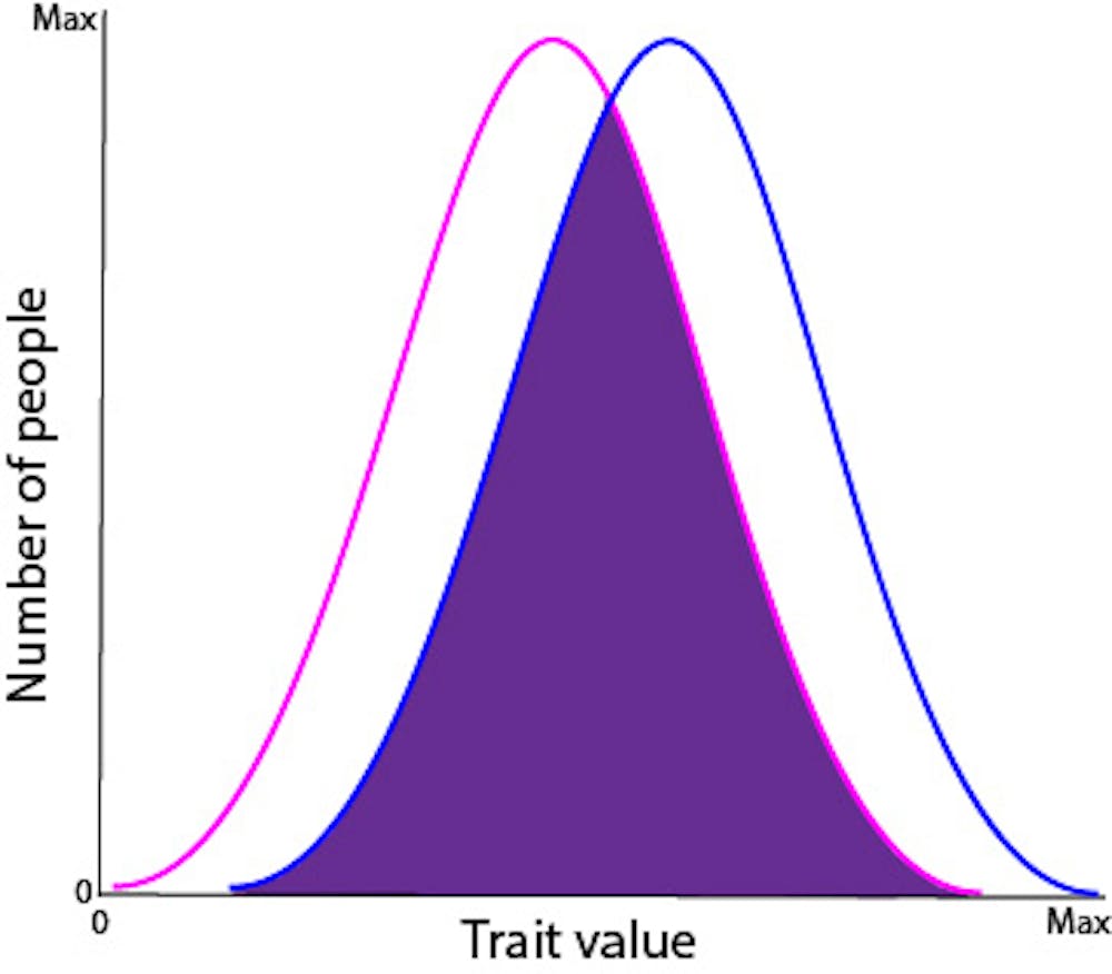
Even when a brain region shows a sex difference on average, there is typically considerable overlap between the male and female distributions. If a trait’s measurement is in the overlapping region, one cannot predict the person’s sex with confidence. For example, think about height. I am 5’7". Does that tell you my sex? And brain regions typically show much smaller average sex differences than height does.
Neuroscientist Daphna Joel and her colleagues examined MRIs of over 1,400 brains, measuring the 10 human brain regions with the largest average sex differences. They assessed whether each measurement in each person was toward the female end of the spectrum, toward the male end or intermediate. They found that only 3% to 6% of people were consistently “female” or “male” for all structures. Everyone else was a mosaic.
Prenatal hormones
When brain sex differences do occur, what causes them?
While prenatal hormones probably cause most brain sex differences in nonhumans, there are some cases where the cause is directly genetic.
This was dramatically shown by a zebra finch with a strange anomaly – it was male on its right side and female on its left. A singing-related brain structure was enlarged (as in typical males) only on the right, though the two sides experienced the same hormonal environment. Thus, its brain asymmetry was not caused by hormones, but by genes directly. Since then, direct effects of genes on brain sex differences have also been found in mice.
Learning changes the brain
Many people assume human brain sex differences are innate, but this assumption is misguided.
Humans learn quickly in childhood and continue learning – alas, more slowly – as adults. From remembering facts or conversations to improving musical or athletic skills, learning alters connections between nerve cells called synapses. These changes are numerous and frequent but typically microscopic – less than one hundredth of the width of a human hair.
Some London taxi drivers do not use GPS – they know the city by heart, a learning process that takes three to four years on average.
Studies of an unusual profession, however, show learning can change adult brains dramatically. London taxi drivers are required to memorize “the Knowledge” – the complex routes, roads and landmarks of their city. Researchers discovered this learning physically altered a driver’s hippocampus, a brain region critical for navigation. London taxi drivers’ posterior hippocampi were found to be larger than nondrivers by millimeters – more than 1,000 times the size of synapses.
So it’s not realistic to assume any human brain sex differences are innate. They may also result from learning. People live in a fundamentally gendered culture, in which parenting, education, expectations and opportunities differ based on sex, from birth through adulthood, which inevitably changes the brain.
Ultimately, any sex differences in brain structures are most likely due to a complex and interacting combination of genes, hormones and learning.
Herbivores face a higher risk of extinction than predators, whether they are mammals, birds or reptiles, according to an extensive study of 24,500 species both living and extinct that was published Wednesday.
The paper, which appeared in Science Advances, suggests herbivores have suffered a higher extinction rate over the past 50,000 years compared to other parts of the food web and the trend continues to this day.
This contradicts the idea, based on anecdotal evidence, that predators are the most vulnerable because they have extensive home ranges and slow population growth rates.
The threat is greatest for reptile herbivores, such as turtles, and large herbivores, like elephants.
"There is so much data out there and sometimes you just need someone to organize it," said Trisha Atwood, an ecologist at Utah State University and the first author of the study.
Researchers first looked at modern day extinction risk patterns among herbivores, omnivores and predators in mammals, birds and reptiles at different levels of the food web.
They performed the same analysis on species from the late Pleistocene epoch, beginning 11,000 years ago for Africa, North America and South America, and 50,000 years ago for Australia
Finally, they examined how body size and position in the food web affected the threat status among 22,166 living species.
The authors wrote that though there are probably several reasons for the trend, certain man made interventions seemed to affect herbivores more than others.
"Invasive vertebrates (e.g., rats), insects (e.g., fire ants), and plants (e.g., Hottentot fig) have all been implicated in the decline and even extinction of several reptiles," they said.
What's more, invasive species, pollution and habitat alteration appeared to affect small herbivorous birds disproportionately.
There are certain exceptions: predators living in marine habitats did face an elevated extinction risk, suggesting they faced existential pressures than their land-dwelling counterparts.
Even as the COVID-19 pandemic cripples the economy and kills hundreds of people each day, there is another epidemic that continues to kill tens of thousands of people each year through opioid drug overdose.
Opioid analgesic drugs, like morphine and oxycodone, are the classic double-edged swords. They are the very best drugs to stop severe pain but also the class of drugs most likely to kill the person taking them.
In a recent journal article, I outlined how a combination of state-of-the-art molecular techniques, such as CRISPR gene editing and brain microinjection methods, could be used to blunt one edge of the sword and make opioid drugs safer.
I am a pharmacologist interested in the way opioid drugs such as morphine and fentanyl can blunt pain. I became fascinated in biology at the time when endorphins – natural opioids made by our bodies – were discovered. I have been intrigued by the way opioid drugs work and their targets in the brain, the opioid receptors, for the last 30 years. In my paper, I propose a way to prevent opioid overdoses by modifying an opioid user’s brain cells using advanced technology.
Opioid receptors stop breathing
Opioids kill by stopping a person from breathing (respiratory depression). They do so by acting on a specific set of respiratory nerves, or neurons, found in the lower part of the brain that contain opioid receptors. Opioid receptors are proteins that bind morphine, heroin and other opioid drugs. The binding of an opioid to its receptor triggers a reaction in neurons that reduces their activity. Opioid receptors on pain neurons mediate the pain-killing, or analgesic, effects of opioids. When opioids bind to opioid receptors on respiratory neurons, they slow breathing or, in the case of an opioid overdose, stop it entirely.
Respiratory neurons are located in the brainstem, the tail-end part of the brain that continues into the spine as the spinal cord. Animal studies show that opioid receptors on respiratory neurons are responsible for opioid-induced respiratory depression – the cause of opioid overdose. Genetically altered mice born without opioid receptors do not die from large doses of morphine unlike mice with these receptors present.
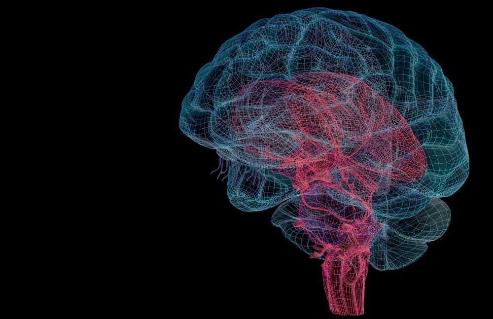
The brainstem is the the red part protruding from the bottom of the brain.
Unlike laboratory mice, humans cannot be altered when embryos to remove all opioid receptors from the brain and elsewhere. Nor would it be a good idea. Humans need opioid receptors to serve as the targets for our natural opioid substances, the endorphins, which are released into the brain during times of high stress and pain.
Also, a total opioid receptor knockout in humans would leave that person unresponsive to the beneficial pain-killing effects of opioids. In my journal article, I argue that what is needed is a selective receptor removal of the opioid receptors on respiratory neurons. Having reviewed the available technology, I believe this can be done by combining CRISPR gene editing and a new neurosurgical microinjection technique.
CRISPR to the rescue: Destroying opioid receptors
CRISPR, which is an acronym for clustered regularly interspaced short palindromic repeats, is a gene editing method that was discovered in the genome of bacteria. Bacteria get infected by viruses too and CRISPR is a strategy that bacteria evolved to cut-up the viral genes and kill invading pathogens.
The CRISPR method allows researchers to target specific genes expressed in cell lines, tissues, or whole organisms, to be cut-up and removed – knocked out – or otherwise altered. There is a commercially available CRISPR kit which knocks out human opioid receptors produced in cells that are grown in cell cultures in the lab. While this CRISPR kit is formulated for in vitro use, similar conditional opioid receptor knock-out techniques have been demonstrated in live mice.
To knockout opioid receptors in human respiratory neurons, a sterile solution containing CRISPR gene-editing molecules would be prepared in the laboratory. Besides the gene-editing components, the solution contains chemical reagents that allow the gene-editing machinery to enter the respiratory neurons and make their way into the nucleus and into the neuron’s genome.
How does one get the CRISPR opioid receptor knockout solution into a person’s respiratory neurons?
Enter the intracranial microinjection instrument (IMI) developed by Miles Cunningham and his colleagues at Harvard. The IMI allows for computer-controlled delivery of small volumes of solution at specific places in the brain by using an extremely thin tube – about twice the diameter of a human hair – that can enter the brain at the base of the skull and thread through brain tissue without damage.
The computer can direct the robotic placement of the tube as it is fed images of the brain taken before the procedure using MRI. But even better, the IMI also has a recording wire embedded in the tube that allows measurement of neuronal activity to identify the right group of nerve cells.
Because the brain itself feels no pain, the procedure could be done in a conscious patient using only local anesthetics to numb the skin. Respiratory neurons drive the breathing muscles by firing action potentials which are measured by the recording wire in the tube. When the activity of the respiratory neurons matches the breathing movements by the patients, the proper location of the tube is confirmed and the CRISPR solution injected.
The call for drastic action
Opioid receptors on neurons in the brain have a half-life of about 45 minutes. Over a period of several hours, the opioid receptors on respiratory neurons would degrade and the CRISPR gene-editing machinery embedded in the genome would prevent new opioid receptors from appearing. If this works, the patient would be protected from opioid overdose within 24 hours. Because the respiratory neurons do not replenish, the CRISPR opioid receptor knockout should last for life.
With no opioid receptors on respiratory neurons, the opioid user cannot die from opioid overdose. After proper backing from National Institute on Drug Abuse and leading research and health care institutions, I believe CRISPR treatment could enter clinical trials in between five to 10 years. The total cost of opioid-involved overdose deaths is about US$430 billion per year. CRISPR treatment of only 10% of high-risk opioid users in one year would save thousands of lives and $43 billion.
Intracranial microinjection of CRISPR solutions might seem drastic. But drastic actions that are needed to save human lives from opioid overdoses. A large segment of the opioid overdose victims are chronic pain patients. It may be possible that chronic pain patients in a terminal phase of their lives and in hospice care would volunteer in phase I clinical trials for the CRISPR opioid receptor knockout treatment I propose here.
Making the opioid user impervious to death by opioids is a permanent solution to a horrendous problem that has resisted efforts by prevention, treatment and pharmacological means. Steady and well-funded work to prove the CRISPR method, first with preclinical animal models then in clinical trials, is a moonshot for the present generation of biomedical scientists.
"We are studying the risk of death faced by our own children."
A new study warns that the annual global death rate from the climate crisis could equal or even exceed current mortality levels from all infectious diseases combined by the end of the century if bold action is not taken.
"We are studying the risk of death faced by our own children," said University of California public policy professor Solomon Hsiang, one of the report's co-authors. "Today's 10-year-old fifth grader will turn 65 in 2075, facing mortality risks from climate change every year of their retirement. Failing to address climate change is not that different from driving your kids around without a seat belt: you are putting their lives at risk."
The study, "Valuing the Global Mortality Consequences of Climate Change Accounting for Adaptation Costs and Benefits," was published in the National Bureau of Economic Research on Monday.
According to Climate Impact Lab, which produced the report, the study "finds that in a world with continued high fossil-fuel emissions, warmer temperatures will rank among the world's most significant public health threats by the end of the century."
As Climate Impact Lab explained:
The study projects that climate change's effect on temperatures could raise global mortality rates by 73 deaths per 100,000 people in 2100 under a continued high emissions scenario, compared to a world with no warming. That level is roughly equal to the current death rate for all infectious diseases—including tuberculosis, HIV/AIDS, malaria, dengue, yellow fever, and diseases transmitted by ticks, mosquitos, and parasites—combined (approximately 74 deaths per 100,000 globally).
"Our data indicate that with the continued growth of greenhouse gas emissions, the temperature effects of climate change are projected to be five times deadlier than recent U.S. flu seasons," said report co-author Michael Greenstone, a University of Chicago economics professor. "In poor hot countries, the heat may be even more threatening than cancer and heart disease are today."
The study also found that the economic cost of addressing the climate crisis will only increase if little or nothing is done to reduce emissions—"emitting one additional ton of CO2 today costs ourselves and future generations a total of $36.6 under a continued high emissions scenario and $17.1 under a moderate emissions scenario."
"Just as countries are impacted in different ways by extreme temperatures today, we find that the trend will continue and perhaps even intensify into the future as adaptation becomes more and more critical to people's survival," said co-author Amir Jina, an assistant professor at the University of Chicago Harris School of Public Policy. "Indeed, some will need to choose between paying a high cost to adapt and death."
Citing the social and economic upheaval sparked by the current Covid-19 pandemic, Jina told the Guardian that the unchecked climate crisis would have far-reaching negative impacts.
"It's plausible that we could have the worst-case scenario and that would involve drastic measures such as lots of people migrating," Jina said. "Much like when Covid overwhelms a healthcare system, it's hard to tell what will happen when climate change will put systems under pressure like that. We need to understand the risk and invest to mitigate that risk, before we really start to notice the impacts."
Trevor Houser, a partner at analyst firm Rhodium Group and another report co-author, sounded a hopeful note, saying that if nations take decisive action now to combat the climate crisis, there's hope that the dire predictions in the study won't come to pass.
"The world can still change course by aggressively reducing emissions, and in doing so has the potential to deliver some of the most significant public health gains in human history," said Houser.
The question of whether ancient life could have existed on Mars centers on the water that once flowed there, but new research published Monday suggests that many of the Red Planet's valleys were gouged by icy glaciers not rivers.
The study in Nature Geoscience, which comes amid a flurry of new Mars missions trying to discover if the now-barren planet ever hosted life, casts doubt on a dominant theory that the planet once had a warm, wet climate with abundant liquid water that sculpted the landscape.
Researchers from Canada and the United States examined more than 10,000 Martian valleys and compared them to channels on Earth that were carved under glaciers.
"For the last 40 years, since Mars's valleys were first discovered, the assumption was that rivers once flowed on Mars, eroding and originating all of these valleys," said lead author Anna Grau Galofre in a statement released by the University of British Columbia.
But these formations come in a huge variety "suggesting that many processes were at play to carve them," she added.
Researchers found similarities between some Martian valleys and the subglacial channels of Devon Island, in the Canadian Arctic, which has been nicknamed "Mars on Earth" for its barren, freezing conditions and hosted NASA space training missions.
The study authors said their findings suggest that some Martian valleys could have been formed some 3.8 billion years ago by meltwater beneath ice sheets, which they said would align with climate modeling predicting that the planet would have been much cooler in its ancient past.
"The findings demonstrate that only a fraction of valley networks match patterns typical of surface water erosion, which is in marked contrast to the conventional view," said co-author Mark Jellinek.
Nature Geoscience noted that understanding climate conditions "in the first billion years of Mars' history is important in determining whether the planet was ever habitable".
The study authors said that icy temperatures could in fact have better supported ancient life.
"A sheet of ice would lend more protection and stability of underlying water, as well as providing shelter from solar radiation in the absence of a magnetic field - something Mars once had, but which disappeared billions of years ago," the University of British Columbia statement said.
The research comes after NASA launched its latest Mars rover, Perseverance, to look for signs of ancient microbial life on the Red Planet.
If all goes to plan, Perseverance will reach Mars on February 18, 2021 and collect rock samples that could provide invaluable clues about whether there was ever past life on Mars.
However, the retrieval and analysis is not expected before the 2030s.
China has also launched its first Mars rover, which should arrive by May 2021.
Supporters of President Donald Trump on Sunday were accused of jeopardizing astronauts' return to Earth.
After SpaceX's Crew Dragon capsule became the first American spacecraft to splashdown in 45 years, a boat flying the Trump flag was spotted within feet of the astronauts.
The boaters were quickly called out on social media for "interfering" with the mission.
Watch the video and read some of the responses below.
The first US astronauts to reach the International Space Station on an American spacecraft in nearly a decade might not come home this weekend as scheduled because of Hurricane Isaias, NASA said Friday.
Bob Behnken and Doug Hurley blasted off from Cape Canaveral on May 30 on board a SpaceX Crew Dragon, and are supposed to splash down off the coast of Florida on Sunday afternoon.
For now, undocking remains scheduled for approximately 7:34 pm (2334 GMT) Saturday, and splashdown at 2:42 pm (1842 GMT) on Sunday.
But NASA said it was keeping a watchful eye on Hurricane Isaias -- a category one storm that battered the Bahamas Friday and was churning toward Florida -- and would make a final call about six hours prior to undocking.
"We don't control the weather, and we know we can stay up here longer –- there's more chow, and I know the space station program has more work that we can do," Behnken told reporters in a press call.
The potential splashdown sites are in the Gulf of Mexico and along Florida's Atlantic coast.
The mission marked the first time a crewed spaceship launched into orbit from American soil since 2011 when the Space Shuttle program ended.
It was also the first time a private company has flown to the ISS carrying astronauts.
The US has paid SpaceX and aerospace giant Boeing a total of about $7 billion for their "space taxi" contracts.
But Boeing's program has floundered badly after a failed test run late last year, which left SpaceX, a company founded only in 2002, as clear frontrunner.
For the past nine years, American astronauts traveled exclusively on Russian rockets Soyuz rockets, for a price of around $80 million per seat.
The crewmates added they were looking forward to going home after two months, if the return trip went ahead Sunday as planned.
"My son is six years old and I can tell from the videos that I get and from talking to him on the phone that he's changed a lot," said Behnken.
The assembly of the ITER fusion reactor began in the south of France this week in what has been called the biggest science project in human history. It is hoped the reactor will be able to produce clean energy using the same process that fuels the sun.
ITER (International Thermonuclear Experimental Reactor) is an international project that hopes to create clean energy from hydrogen fusion, the same process that occurs naturally in the heart of the sun.
Fusion will be obtained through a mixture of two hydrogen isotopes, heated to a temperature of around 150 million degrees.
"In this space we are going to have a machine in the heart of which a small sun will burn, to put it very simply. This small sun will generate energy. We will use that energy to create electricity,” ITER spokesperson Robert Arnoux told AFP.
A long sought-after alternative to fossil fuels such as coal and gas, hydrogen fusion generates no long-term waste, says ITER, while the fuels needed to create fusion are found in seawater and lithium, and so readily available.
ITER should start producing energy at the end of 2025 or early 2026 but only on an experimental basis. It will be a number of years before it is capable of supplying usable electricity.
"The vision I have, the one I think is most realistic, is that we'll have to wait until about 2060 before we can connect the first fusion-powered generator to a power grid,” said Bernard Bigot, ITER director general.
The ITER project began in 2006 with a treaty bringing together 35 countries, including those in the EU, the UK, Switzerland, Russia, China, India, Japan, South Korea and the US.
Some environmental groups have criticised the ITER project dubbing it a ‘money pit’ and a ‘scientific mirage’.
The project is already five years behind schedule and three times over the initial budget at a cost of almost €20 billion.